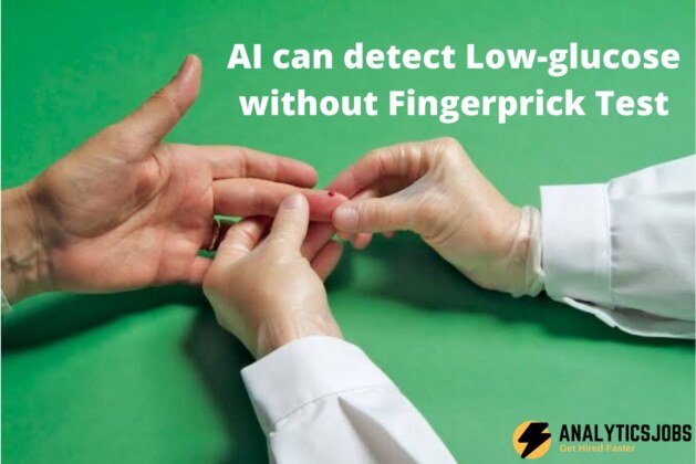
Tracking sugars in the blood is essential for both healthy people and diabetic patients. Present techniques to measure glucose involves needles and repeated fingerpicks. Fingerpicks can usually be painful, deterring patient compliance. AI can detect low-glucose with no fingerprick test.
A new technique created by researchers at the University of Warwick uses the latest results of Artificial Intelligence to identify hypoglycaemic events from raw ECG indicators, via wearable receptors or sensors. The technology works with an 82% reliability and may change the need for invasive fingerprick tests with a needle, which may be especially helpful for pediatric age patients.
New technology for detecting lower glucose levels via ECG making use of a non-invasive wearable sensor, that with the latest Artificial Intelligence is able to detect hypoglycaemic events from raw ECG indicators has been created by researchers from the University of Warwick Dr. Leandro Pecchia with the new technology.
Continuous Glucose Monitors (CGM) are available by the NHS for hypoglycemia detection (sugar levels into derma or blood). They calculate glucose in interstitial material making use of an invasive sensor with a little needle, which transmits data and alarms to a display device. In various cases, they need calibration twice a day with invasive finger-prick blood glucose levels test.
2 pilot studies with wholesome volunteers found the average sensitivity and specificity more or less 82% for hypoglycemia detection, which is equivalent to the existing CGM efficiency, although non-invasive.
Fingerpicks are never pleasant and in some cases are particularly troublesome. Taking fingerpick during the evening definitely is uncomfortable, particularly for patients in the pediatric age. Our innovation consisted of utilizing artificial intelligence for automatic detecting hypoglycemia via not many ECG beats. This is relevant because ECG may be detected in any condition, including sleeping.
The figure displays the output of the algorithms over time. The green line indicates normal glucose levels, and the red line represents the low glucose levels. The horizontal line symbolizes the 4mmol/L glucose value, which is regarded as the major threshold for hypoglycaemic events. The grey area that involves the constant line reflects the measurement error bar.
The Warwick design highlights how the ECG switches in each subject during a hypoglycaemic event. The figure below is an exemplar. The solid lines represent the average heartbeats for 2 different subject matter when the glucose level is natural (green line) or perhaps minimal (red line). The green and red shadows stand for the regular deviation of the heartbeats across the mean.
A comparison highlights that these 2 subjects have various ECG waveform switches during hypo events. Particularly, Subject 1 presents a visibly longer QT interval in the course of hypo, while subject two doesn’t.
The vertical bars stand for the relative value of each ECG wave in finding out whether a heartbeat is classified as hypo or even normal.
From these bars, a skilled clinician views that for Subject 1, the T wave displacement influences distinction, reflecting that once the subject is within hypo, the repolarisation on the ventricles is slower.
In Subject 2, the most crucial components of the ECG are rising of the T wave and the P-wave, hinting that when this particular subject is in hypo, the depolarization of the threshold and the atria for ventricular activation are affected. This may influence the following clinical interventions.
This outcome is feasible because the Warwick AI model is taught with each subject’s own details. Intersubjective differences are very important, that training the system using cohort data wouldn’t give the exact same results. Likewise, personalized therapy based mostly on the system of ours might be better compared to current approaches.
AI can detect Low-glucose without Fingerprick Test
Analytics Jobs


